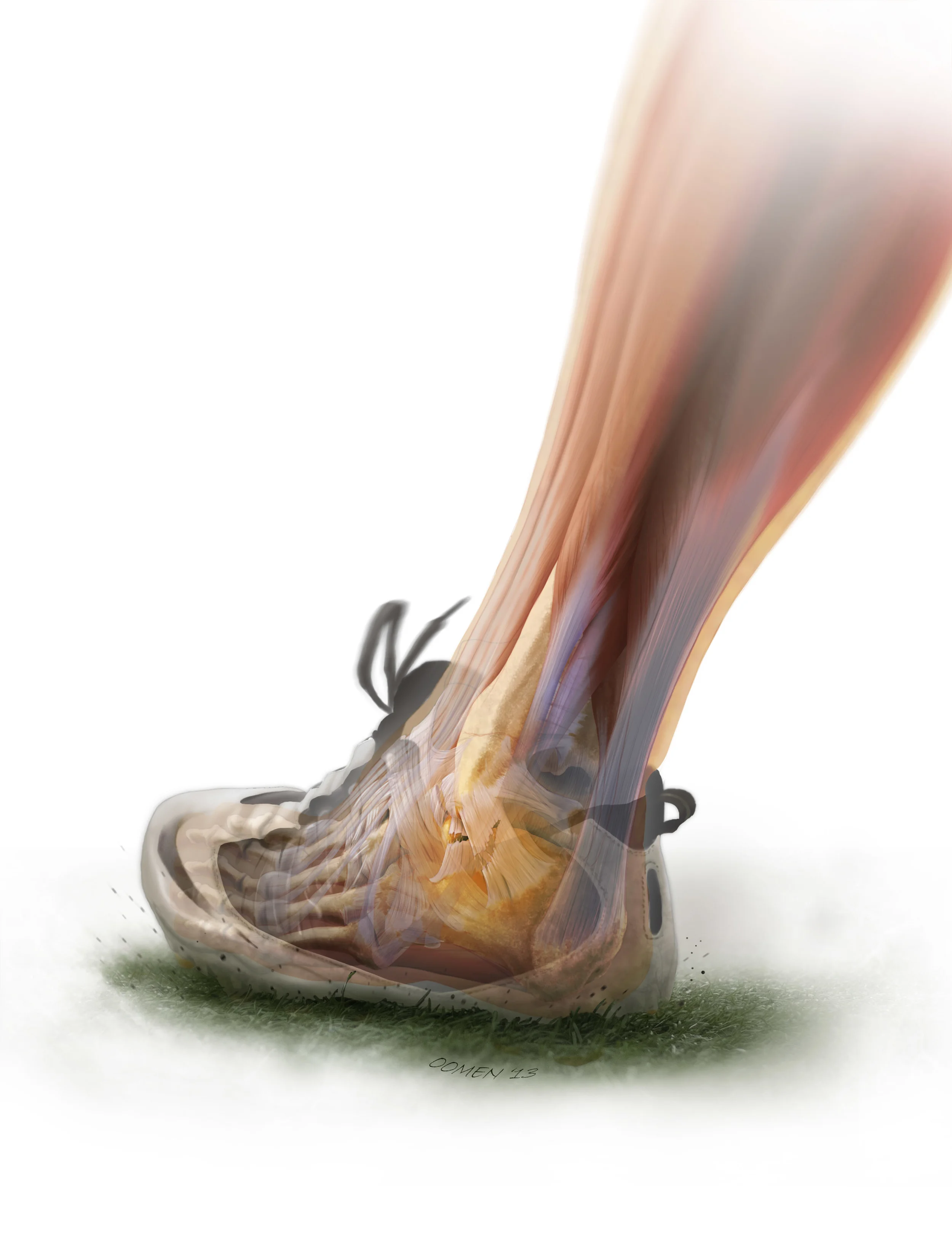


Dynamic illustration showing the classic lateral ankle sprain.
One in a series illustrating a technique for maximizing cerebral protection in repair of ascending aortic aneurysm.
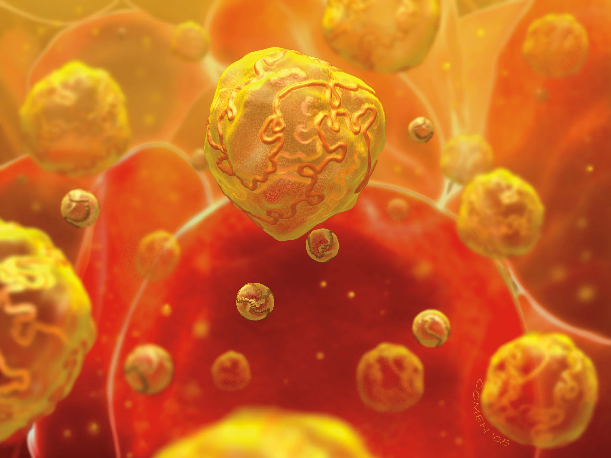
This illustration is an old favorite. It was used a journal cover for an edition examining the relationship between hypertension and low/high density lipoproteins.
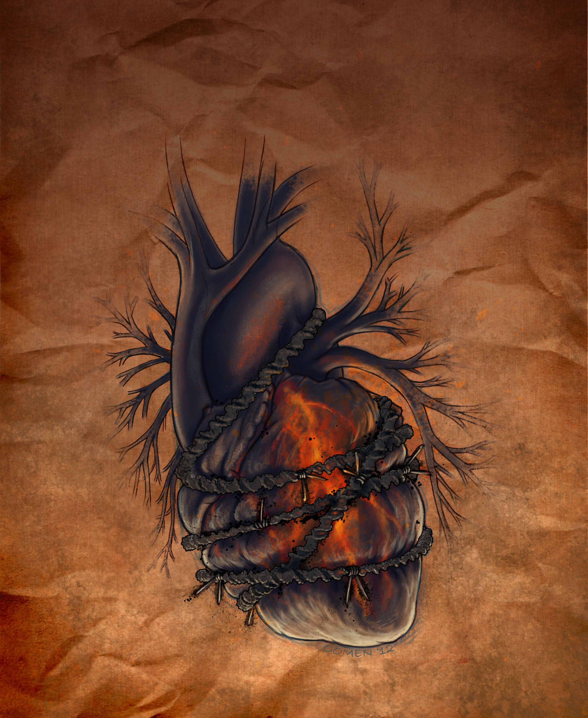
This illustration was done for the cover of the Journal of Biological Chemistry. It was intended to illustrate gene deficiencies that predisposed patients cardiac tissue death when being treated with Doxorubicin, a common chemotherapeutic.
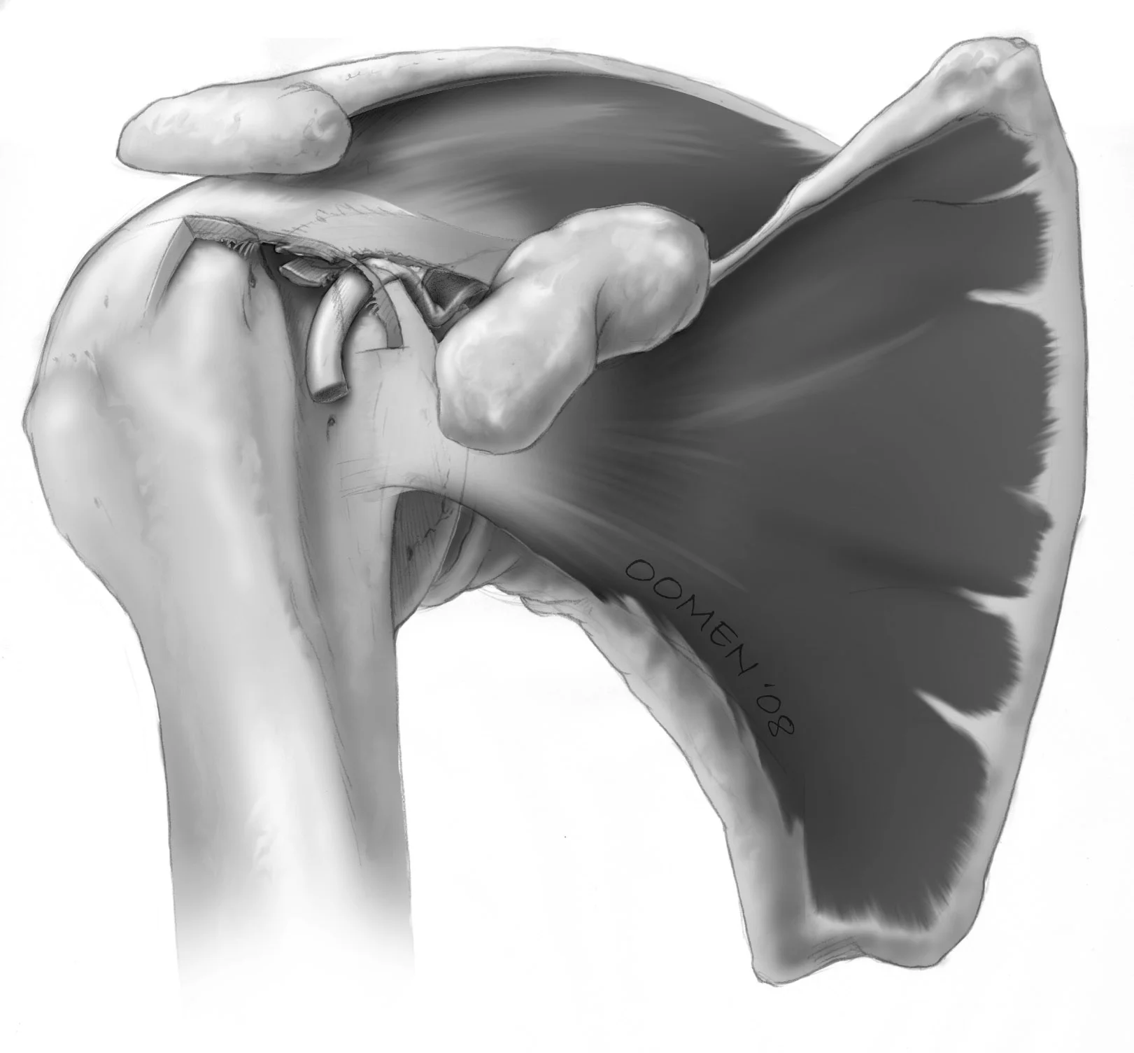
This illustration depicts a pulley rupture, where torn supporting ligaments in the joint capsule of the should allow the biceps tendon to flip out of the intertubercular groove - like a broken pulley.
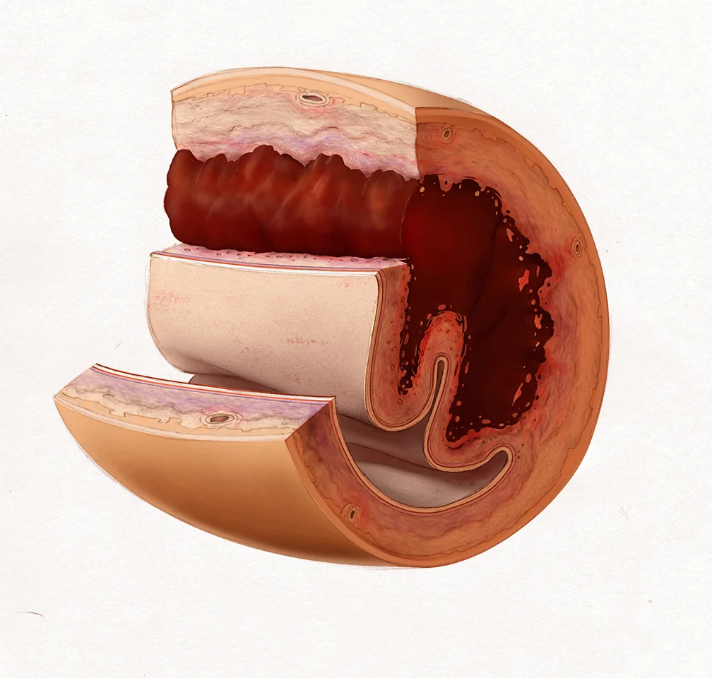
This image illustrates the key pathological features of sudden pregnancy related coronary artery disease, including a large mural thrombus and smooth muscle proliferation.
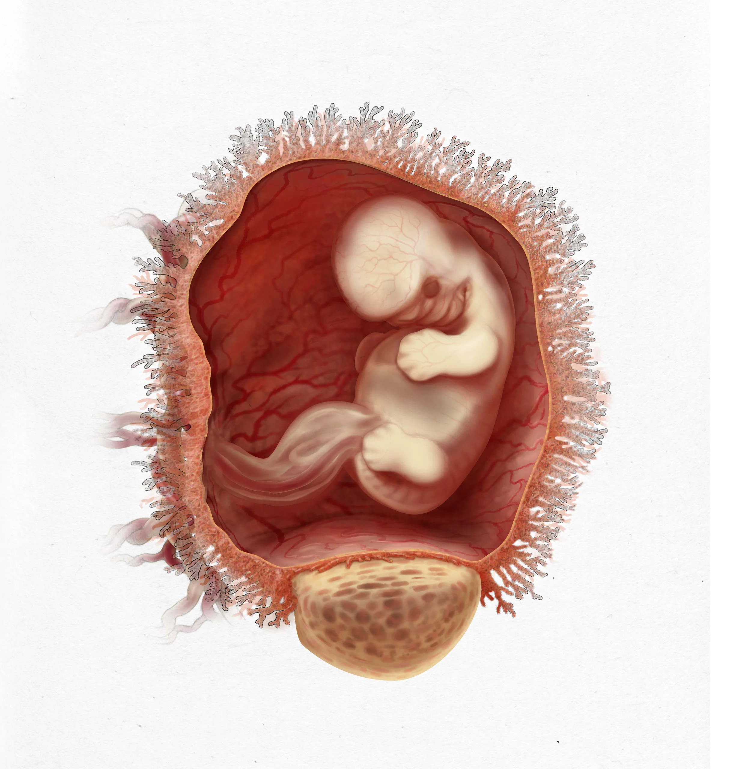
Image depicting a 46 day old human embryo.
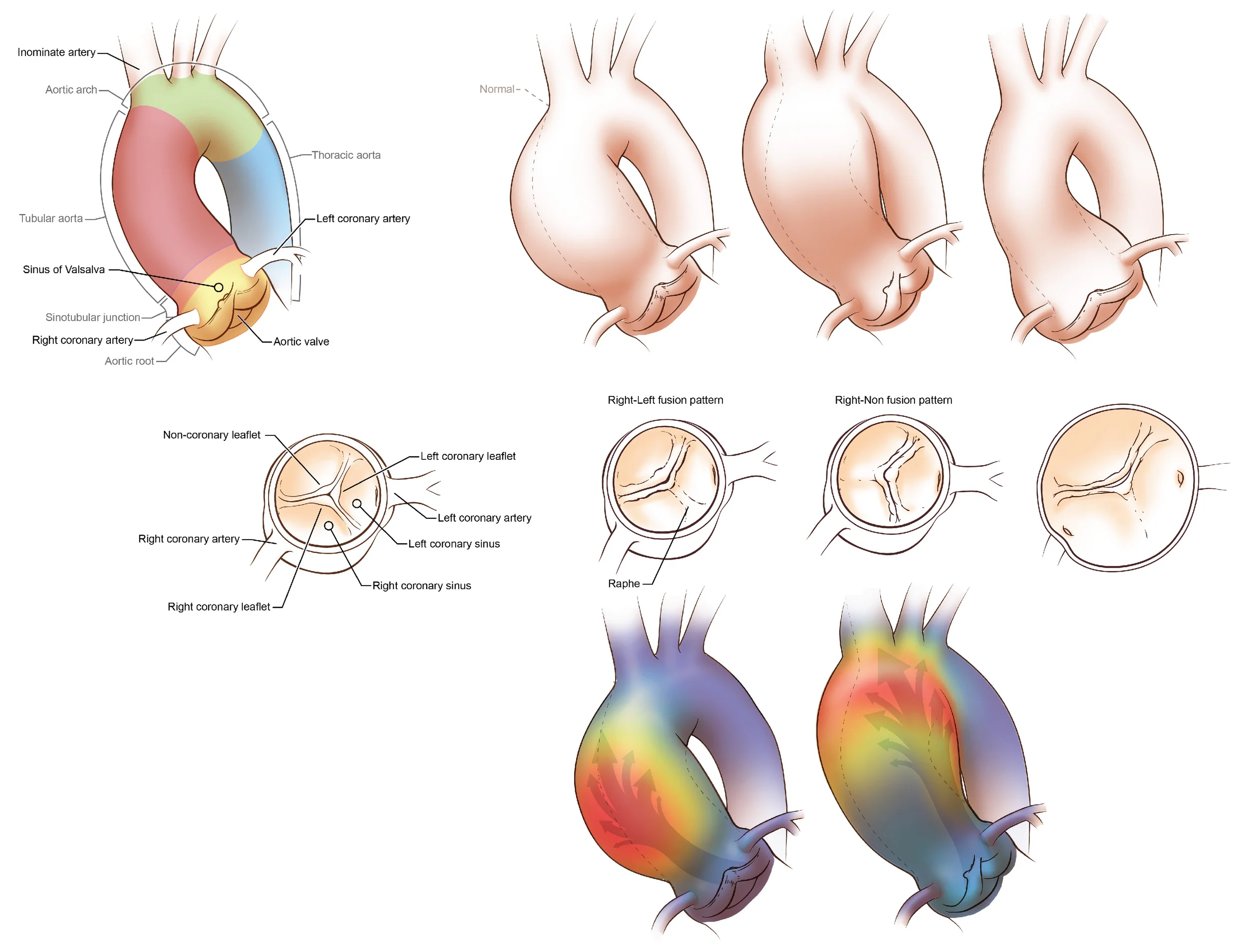
This illustration, originally done for the New England Journal of Medicine, shows the relationship between different leaflet configurations in bicuspid aortic valves, and the ejection fraction each configuration causes. The ejection fractions impose abnormal shear stress on the aortic wall causing different patterns of aneurysm.
Illustration showing the path of the posterior cutaneous nerve of the forearm and how it might relate to lateral epicondylitis.
Cover for the Journal of Biological Chemistry depicting pulmonary fibrosis.
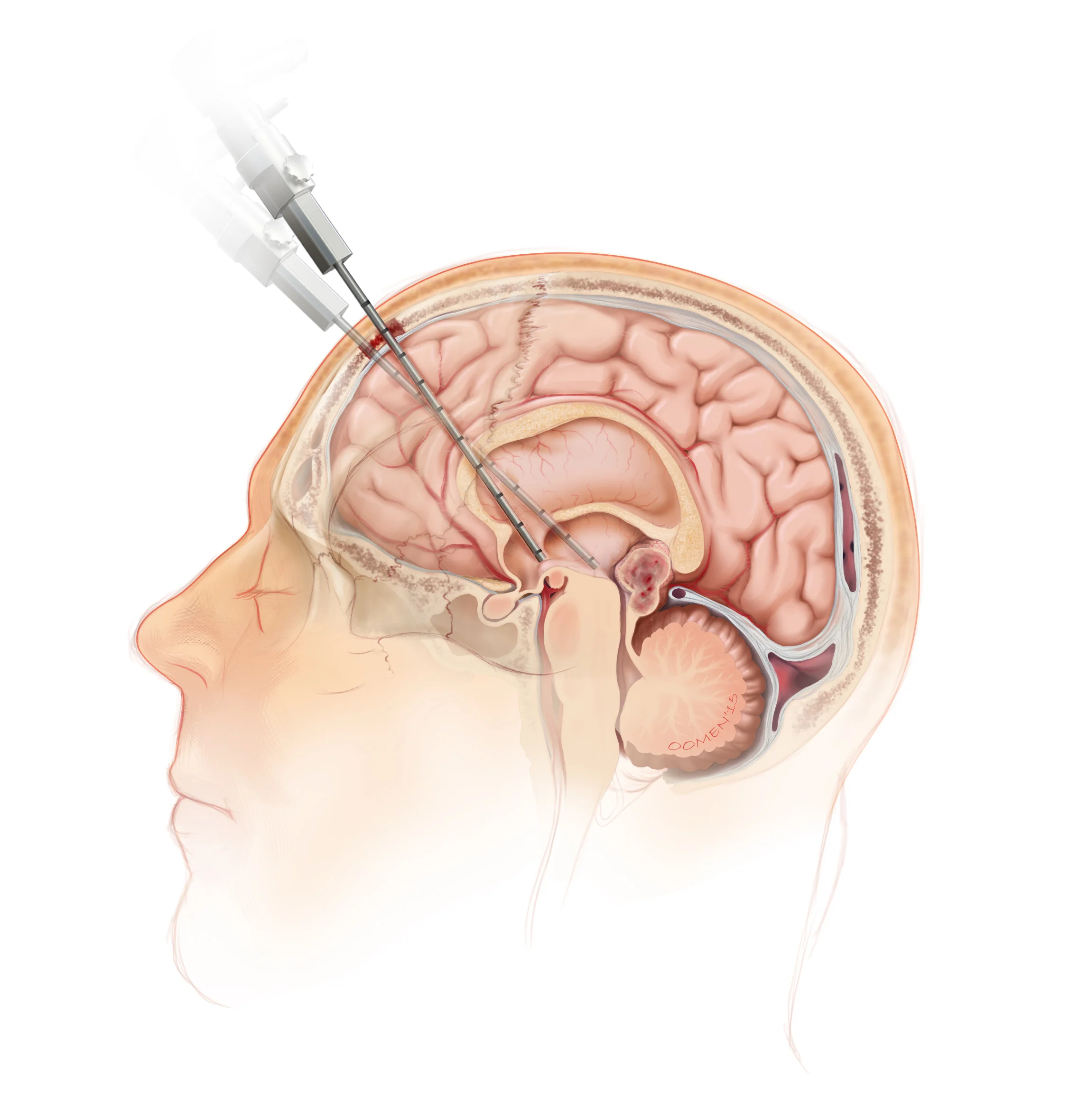
Illustration demonstrating the approach for endoscopic transventricular access to pineal tumours in the third ventricle.
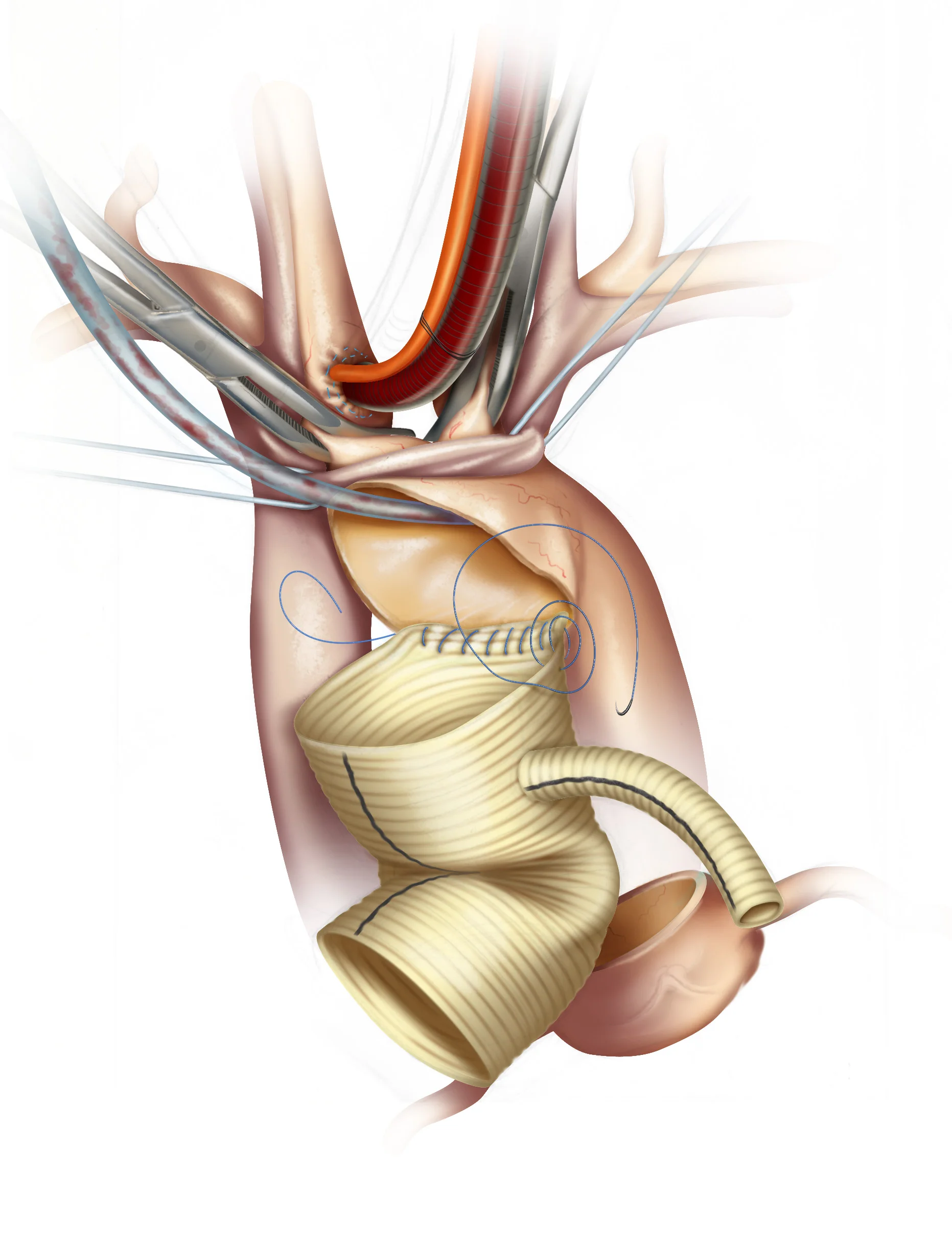
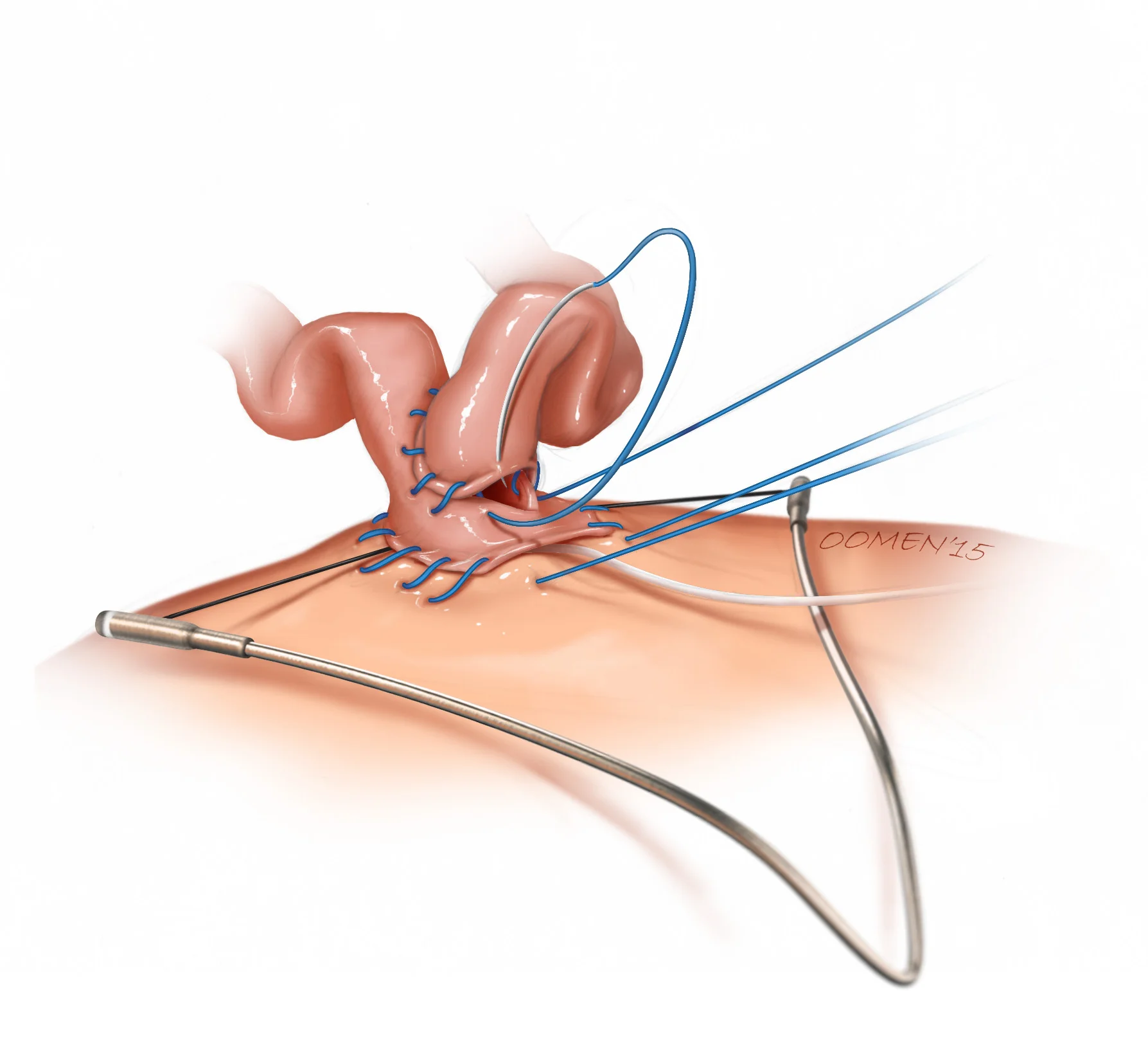
This is one image in a large sequence describing different methods for grafting small-diameter vessels to the aorta during CABG. This image also demonstrates the use of a Heartstring device for hemostasis during the procedure.
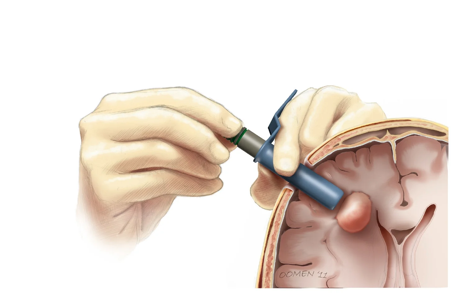
This image was one of in a series demonstrating the neurosurgical use of conventional tube retractors to access lesions within the brain.